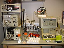
 QUANTIFICATION:
1 Estimation of Glycyrrhizic Acid by HPTLC.
(a) Sample preparation :
- 0.25 g dry extract is taken in 50 ml 1N HCL and refluxed for four hours. After cooling to room temperature, it is extracted with 20 ml chloroform 5 times. The combined chloroform extract, after washing with water and filtration is evaporated at maximum temperature of 300 C and residues is dissolved in chloroform-methanol (1:1) and make up to 25 ml.
(b) Sample Application :
- Bandwise with Linomat 2 , 3 and 4 ul of standard and sample solution, band length 6 mm, distance between bands 4mm.
(c) Chromatography :
- Two step chromatogram development :
1st Run :- Chloroform-Acetone 9:1
2nd Run :- Chloroform-Diethyl Ether-Formic Acid 80:15:1
(d) Quantitative evaluation :
- Scanning by absorbance at 254 nm (Hg lamp) monochromator band width 10 nm. Slit dimension 4 x 0.3, evaluation via peak height or peak area. 1ul of the calibration standard solution contains 400 ng glycyrrhetinic acid corresponding to 700 ng glycyrrhizic acid in the extract.
2 HPTLC Method For Estimation Of Curcuminoids From Curcuma Longa.
Curcuma longa (Zingiberaceae), commonly called Haldi, is a well-known plant drug in Ayurvedic and Unani It has been used for the treatment of various diseases and disorders particularly for urticaria, skin allergy, viral hepatitis, inflammatory condi-tions of joints, sore throat, and for wound.Curcumin, demethoxycurcumin and bis-demetho-xycurcumin, three major pharmacologically important curcuminoids, have been isolated from C. longa : and has been shown to possess anti-oxidant, anti-inflammatory, anti-carcinogenic, anti-mutagenic, anti-fungal, anti-viral and anti-cancer ..Methods, so far available for the determination of these alkaloids, are very cumbersome and time-consuming and also not economically viable. Therefore it was thought worthwhile to develop a simple and high-precision HPTLC method for simultaneous analysis of curcumin, demethoxy curcumin and bis-demethoxycurcumin occurring in roots of C. longa.
Rf values by HPTLC and linear regression equa-tions for the determination of curcumin, demethoxycurcumin and bis-demethoxy-curcumin.
Compound Rf value
Curcumin 0.67
Demethoxycurcumin 0.47
Bis-demethoxy-curcumin 0.29
Chromatographic conditions:
Chromatography was performed on glass-backed silica gel 60 GF254 HPTLC layers (20 x 20 cm,
300 μm layer thickness).
Methanolic solutions of samples and standard compounds curcumin, demethoxycurcumin and bis-demethoxycurcumin of known concentrations were applied to the layers as 7 mm-wide bands positioned 15 mm from the bottom and 20 mm from the side of the plate,
Mobile Phase: chloroform: methanol (48:2, v/v) for one hour.
25 ± 50C and 50 % relative humidity. Quantitative analysis of the compounds was done by scanning the plates using,
Slit width: 6 x 0.45 mm,
Wavelength (λmax): 425 nm.
In order to prepare calibration curves, stock solutions of curcumin, demethoxycurcumin and bis-demetho-xycurcumin (1 mg/5 mL each) were prepared and various volumes of these solutions were analyzed by HPTLC exactly as described above. Then calibration curves of peak area vs. concentration were prepared.
Results and Discussion:
The wave length of 425 nm was found to be optimal for the highest sensitivity. The calibration curves for the alkaloids curcumin, demethoxy-curcumin and bis-demethoxycurcumin were linear in the range 100-1,000 ng
3 HPTLC method for estimation of charantin:
Diabetes mellitus has recently been identified by Indian Council of Medical Research (ICMR) as one of the refractory diseases for which satisfactory treatment is not available in modern allopathic system of medicine and suitable herbal preparations are to be investigated
Momordica charantia Linn. Cucurbitaceae is a well known to possess antihyperglycemia, anticholesterol, immunosuppressive, antiulcerogenic, anti seperma -togenic and androgenic All the three selected polyherbal formulations contain karela as one of the plants. The reference standard charantin had to be isolated, purified and the structure authenticated by various spectral analysis. There are no titrimetric, colorimetric, spectrophotometric or chromatographic methods available for quantitative estimation of charantin in different marketed antidiabetic PHFs. Therefore an attempt has been made to develop a HPTLC method because this method is fast, precise, sensitive and reproducible with good recoveries for standardization of polyherbal formulations.
Chromatographic conditions:
Chemicals: Analytical grade chloroform, benzene, methanol, formic acid ethyl acetate aluminium.
Stationary Phase: Plates pre-coated with silica gel 60 F 254 (10x 10 cms, 0.2 mm thick).
Rf Value: 0.32 was visible and scanned under 536 nm.
Wavelength (λmax): 536 nm.
Procedure:
One milligram of working standard charantin was dissolved in 100 ml of chloroform to yield stock solution of 100μg/ml concentration. Calibration curve from 20-600 ng /spot was prepared and checked for reproducibility, linearity and validating the proposed method. The correlation coefficient, coefficient of variance and the linearity of results were calculated.
Sample preparation:
200 mg of PHFs were taken and extracted in 10 ml of chloroform then the chloroform extract was filtered through Whatmann no. 42 filter paper. The final volume of the extract was made to 10ml with chloroform in volumetric flask. The small and big karela were dried under shade and finely powdered. From that 50 mg of fine powder was taken and extracted by chloroform and filtered dried extract the volume make up to 2 ml with chloroform.
Method Specifications:
Silica gel 60 F254 precoated plates (10 x 10 cm) were used with benzene: methanol (80:20) as solvent system.
plates were developing up to 8 cms. The plates were sprayed with 10% sulphuric acid in alcohol and the reagent was prepared freshly, heated at 1300 C for 2-3 min and brought to room temperature.
HPTLC chromatogram of standard Charantin:
Results and Discussion:
Standard charantin showed single peak in HPTLC chromatogram. The calibration curve of charantin was obtained by spotting standard charantin on HPTLC plate. After development the plate was scanned at 536 nm the calibration curve was prepared by plotting the concentration of charantin versus average area of the peak. The amount of charantin was computed from calibration curve and calibration curve was shown in Fig. 98.89.
The lowest detectable limit of charantin in different formulations was found upto 20 ng/spot.
4 HPTLC method for estimation Sennosides:
Cassia angustifolia (Family: Caesalpiniaceae), popularly known as senna, is a valuable plant drug in ayurvedic and modern system of medicine for the treatment of constipation Sennoside A and B are the two anthraquinone glycosides that are responsible for purgative action of senna. A variety of poly-herbal formulations containing senna leaves are available in India to relief constipation and allied troubles. Senna is a strong purgative that should be taken in proper dosage otherwise it may lead to gripping and colon problem Different analytical techniques, viz, thin layer chromatography, spectrophotometry, column chromatography have been reported in the literature for estimation of sennosides .
Chromatographic conditions:
Stationary Phase:
Chromatography was performed on glass-backed silica gel 60 GF254 (20 cm x 20 cm; 0.30 mm layer thickness)
Samples and standard compounds 1 and 2 of known concentrations were applied as 8 mm wide bands with nitrogen flow providing delivery speed 150 ηl/s from the syringe. These parameters were kept constant through out the analysis.
25 ± 2oC and 60% relative humidity
Slit width: 6 x 0.45 mm.
Wave length (λmax): 350 nm.
Analytical standards of sennoside A and B were obtained from Solvents (methanol, 2-propanol, ethyl acetate, formic acid) used in entire study.
Preparation of standard solutions:
Standard solutions of sennoside A and sennoside B (1 mg\5 ml) were prepared in methanol.
Extraction of samples:
Each 4 g of different branded formulation was sonnicated with 70% methanol (3 x 20 ml) for about 45 min. Then the extract was filtered in a Buchner funnel using Whatman No. 1 filter paper and was concentrated under vacuum in a rotary evaporator at 50ºC, redissolved in methanol and finally reconstituted in 20 ml methanol prior to HPTLC analysis.
Samples and standard compounds 1 and 2 of known concentrations were applied as 8 mm wide bands with nitrogen flow providing delivery speed 150 ηl/s from the syringe. These parameters were kept constant through out the analysis.
Rf values by HPTLC for the determination of sennoside A and B
Compound Rf value
Sennoside A 0.84
Sennoside B 0.63
Sennoside A and B in selected herbal formulations
Herbal formulations Sennoside A
(in mg/g of formulation) Sennoside B
(in mg/g of formulation)
Formulation-1 2.50 25.90
Formulation-2 2.30 12.70
Formulation-3 2.10 2.60
Formulation-4 1.80 1.87
Formulation-5 1.62 1.85
Formulation-6 1.58 1.72
Formulation-7 1.40 4.53
Formulation-8 1.20 1.50
Formulation-9 0.92 1.50
Formulation-10 0.91 1.40
Results and Discussions:
Scan (at 350 nm) showing the separation of sennoside A and B in the extract of a laxative formulation and limit of quantification of sennoside A and B were determined to be 0.05 and 0.25 μg/g.
The result showed that the relative amount of sennoside A and B in formulation-1 was highest and in formulation-10 it was lowest. Theresult reflected a 2.75 times higher concentration in formulation-1 compared to that of formulation-10. The minimum content of sennoside B (considering the highest content as 100%) was shown only by 5.41% in case of formulation-10 and less than 17.5% in case of formulation number ‘3’ to ‘9’.
Sennoside A and B was found in the concentration range 200-1000 ng. Regression analysis of the experimental data points showed a linear relationship with excellent correlation coefficient(r) of sennoside A and sennoside B of 0.991 and 0.997, respectively The average recovery rate was 95% for sennoside A and 97% for sennoside B.
5 HPTLC method for estimation of glucosamine :
Chromatographic conditions:
Stationary Phase: Glucosamine was separated from the plant extracts on a silica gel 60 F254
Chemicals: HPTLC plate using a saturated mixture of 2-propanol–ethyl acetate–ammonia solution (8%) (10:10:10, v/v).
wave length (λmax) : 415nm.
The plates were developed vertically up to a distance of 80 mm. For visualization, the plate was dipped into a modified anisaldehyde reagent and heated at 120 °C for 30 min in a drying oven. Glucosamine appeared as brownish-red chromatographic zones on a colourless background.
Result:
The relative standard deviations for repeatability and intermediate precision were between 4.9 and 8.6%. Moreover, the method was found to be accurate, as the two-sided 95% beta-expectation tolerance interval did not exceed the acceptance limits of 85 and 115% on the whole analytical range (800–1200 ng of glucosamine).
6 HPTLC method for estimation of trans-Resveratrol :
Ttrans-resveratrol is obtain from Polygonum cuspidatum root extracts . [(E)-5-[2-4(hydroxyphenyl)ethenyl]-1,3- benzenediol] is a naturally occurring polyphenolic compound belonging to a group called stilbenes, found in grapes, peanuts and other plants.2) trans-Resveratrol is a strong antioxidant and reported to have a protective effects against atherosclerosis, coronary heart disease, postmenopausal problems, inhibits platelet aggregation and a broad spectrum of degenerative diseases and also possess cancer chemopreventive properties. The roots of Polygonum cuspidatum, (Polygonaceae).
Chromatographic conditions:
Stationary Phase : Silica gel 60F-254.
Mobile phase: Eluted with chloroform–ethylacetate–formic acid (2.5 : 1 : 0.1).The plates were prewashed by methanol and activated at 60 °C for 5 min prior to chromatography. The length of chromatogram run was 8 cm.
Wave length (λmax): 313nm.
Result:
A good linear regression relationship between peak areas and the concentrations was obtained over the range of 0.5—3.0 mg/spot with correlation coefficient 0.9989.
The limit of detection and quantification was found to be 9 and 27 ng/spot at Rf value
of 0.40.
The spike recoveries were within 99.85 to 100.70%.
The RSD values of the precision in the range 0.37— 1.84%.
7 HPTLC method for estimation of Phyllanthin and Hypophyllanthin in Phyllanthus Species.
The genus Phyllanthus (Euphorbiaceae) contains 550–750 species in 10–11 subgenera that are distributed in all tropical regions of the world from Africa to Asia, South America and the West Indies. Phyllanthus amarus is the most widespread species and is typically to be found along roads and valleys, and on riverbanks and near lakes in tropical areas. Other species found in India are P. fraternus, P. urinaria, P. virgatus, P.maderaspatensis and P. debilis. The genus Phyllanthu has a long history of use in the treatment of diabetes, intestinal parasites and liver, kidney and bladder problems. P. amarus is highly valued in the treatment of liver ailments and kidney stones and has been shown to posses anti-hepatitis B virus surface antigen activity in both in vivo and in vitro studies.
The major lignans of the genus, namely, phyllanthin (1) and hypophyllanthin (2), have been shown to be anti-hepatotoxic against carbon tetrachloride- and galactosamine-induced hepatotoxicity in primary cultured rat hepatocytes . Thus, an appropriate analytical procedure for the quantitative determination of these lignans in different Phyllanthus species is of considerable importance.
Mobile Phase:
The phase finally chosen, namely, hexane:acetone:ethyl acetate (74:12:8, v/v/v) gave good resolution of phyllanthin (1) and hypophyllanthin (2) from other closely related lignans.
Rf values: 0.24 and 0.29, respectively.
Sample Collection
All the Hypericum species were collected from various locations in the region of Pinar del Río, Cuba. Specimen samples were all collected at blossoming time with most of the flowers opened from April to November in 2000 and 2001. The samples were dried at room temperature for seven days and powdered. Voucher specimens of H. tetrapetalum,
H. nitidum and H. styphelioides.
Sample preparation
Powered samples (0.5 g) were extracted with 10 ml of methanol using an ultrasonic bath for 10 min. For and HPTLC analysis the extracts were first filtered.
Standard preparation
Commercial hypericin formulation tablets were used. Three tablets (0.9 g) equivalent to 2.7 mg of hypericine were powdered and extracted with 10 ml of methanol using an ultrasonic bath for 10 min.
Extraction procedure.
Air-dried leaves (1 g) of P. amarus were separately extracted with either hexane, chloroform, ethyl acetate or methanol. In each case, the extraction was carried out three times with 10 mL of solvent for 10 h at room temperature (25 ± 5°C), and the solvent was removed from the combined extract under reduced pressure to yield the respective crude residue. In order to determine the appropriate extraction solvent, each of the crude residues was dissolved separately in 1 mL of methanol and the contents of the two lignans in each sample was determined by HPTLC. The maximum content of lignans 1 and 2 was obtained by extracting with methanol .
Analytical procedure:
Air-dried leaves (1 g) of different Phyllanthus species were extracted separately at room temperature (25 ± 5°C) with methanol (3 × 10 mL; 10 h for each extraction), and the combined extracts were filtered, dried under vacuum and made up to 1 mL with methanol prior to HPTLC analysis.
Result:
The content of the latter (0.858%) was reportedly higher than that of the former (0.709%).
It was observed that the reported higher concentration of 2 was due to other lignans present at the same Rf as that of hypophyllanthin. The phase finally chosen, namely, hexane:acetone:ethyl acetate (74:12:8, v/v/v) gave good resolution of phyllanthin (1) and hypophyllanthin (2) from other closely related lignans (Fig. 1).
8 Estimation Of β-Asarone In Acorus Calamus Dry Extract By HPTLC.
1) Chromatographic condition:
Mobile phase : Toluene-Ethyl Acetate (93:7).
Wave length : 290nm.
2) Standard and Sample Preparation :
a) Standard Preparation :
A 60 μg/ml solution of β-asarone reference standard is prepared in toluene.
b) Sample Preparation:
About 1 g of Acorus calamus dry extract is accurately weighed and dissolved in 5-7 ml of toluene, sonicated for about 2 minutes and volume made upto 10 ml. The solution is filtered.1 ml of this solution is further diluted to 50 ml and used for further analysis.
3) Procedure :
- Four spots of the standard preparation and four spots of the sample preparation are applied about 1 cm form the edge of the TLC plates.
- The plate is developed upto 9 cm in the mobile phase, dried at room temperature and scanned.
4) Results and discussions :
- Under the chromatographic conditions described above the Rf & β- asarone is calculated. The chromatogram of standard and sample are studied on following basis.
ii. Calibration curves
iii. Inter-day coefficient of variation for analysis.
iv. Average recovery.
The method gives the best resolution of β-asarone from other constituents of Acorus calamus. Thus this newly developed HPTLC method is quick and reliable for quantitative monitoring of β-asarone in Acorus calamus and in herbal preparations containing Acorus calamus.


0 comments:
Post a Comment